When a doctor evaluates a patient’s swollen joint, he or she may recommend performing a procedure called joint aspiration, or arthrocentesis, immediately. Other times the procedure will be scheduled for later, and the patient may need to make preparations.
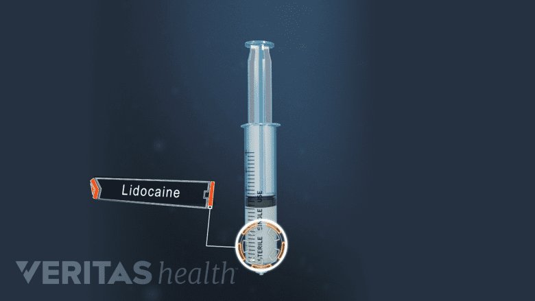
Joint aspiration, can usually be performed quickly and with only local anesthetic such as lidocaine.
Who will perform this procedure? See Arthritis Treatment Specialists
This page reviews basic preparations and provides a step-by-step description of arthrocentesis. The aspiration of excess fluid from a bursa (in cases of bursitis) involves similar preparations and steps.
In This Article:
- What Is Arthrocentesis (Joint Aspiration)?
- The Joint Aspiration Procedure
- Arthrocentesis Recovery and Potential Risks
- Diagnosis through Synovial Fluid Analysis
Preparing for Joint Aspiration (Arthrocentesis)
A needle and syringe are used to aspirate excess fluid from a joint. A local anesthetic may be used to limit pain and discomfort. Before the procedure takes place, a patient should tell the doctor if they:
- Take any medications, including over-the-counter medicines and herbal supplements
- Are allergic to any medications, latex, or anesthesia
- Have any kind of infection or a blood or bleeding disorder
- Are or could be pregnant
Some patients who take blood thinners or anticoagulants may be asked to stop taking these medications in the days leading up to arthrocentesis. Other patients may continue to take these medications but will need to give the aspirated joint a little extra care, such as applying direct pressure to the needle site and resting the joint for a little longer than normal.
Patients who will undergo general anesthesia and those whose joint fluid will be tested for glucose levels may need to fast before arthrocentesis. (Most individuals do not need to fast.)
Patients may also want to ask their doctors if their ability to drive or do other essential tasks will be affected in the hours following the procedure.
Arthrocentesis Step-by-Step
When performed in the doctor’s office with a local anesthetic, arthrocentesis or bursal aspiration can take just 5 to 10 minutes. The procedure may take longer if medical imaging (to ensure accurate placement of the needle) or general anesthesia is used.
The steps to arthrocentesis with aspiration are described below.
- The patient is asked to sit or lie down in a position that is comfortable and gives the doctor easy access to the joint. For example, when aspirating the knee, the patient is asked to recline with the leg relaxed and straightened or at a slight angle. If the knee is at an angle, a towel may be put underneath the knee for support.
- The doctor will use a pen to mark the spot where the needle puncture is to be made.
- The skin around the needle puncture site is thoroughly cleaned.
-
Medical imaging, such as ultrasound, fluoroscopy, or computed tomography (CT), may be used to locate and visualize the affected joint capsule (or bursa). An image of the needle’s target will be projected on a screen for the doctor to see.
While the use of medical imaging is not always necessary, it may help the procedure go more smoothly and yield more successful results.1Sibbitt WL Jr, Kettwich LG, Band PA, Chavez-Chiang NR, DeLea SL, Haseler LJ, Bankhurst AD. Does ultrasound guidance improve the outcomes of arthrocentesis and corticosteroid injection of the knee? Scand J Rheumatol. 2012 Feb;41(1):66-72. doi: 10.3109/03009742.2011.599071. Epub 2011 Nov 21. PMID: 22103390. DOI: 10.3109/03009742.2011.599071
Fluoroscopy or commuted tomography (CT) is typically used if the joint or bursa being aspirated is deep below the skin’s surface, such as the shoulder hip or shoulder joint.2Seidman AJ, Limaiem F. Synovial Fluid Analysis. [Updated 2019 Jan 22]. In: StatPearls [Internet]. Treasure Island (FL): StatPearls Publishing; 2020 Jan-. Available from: https://www.ncbi.nlm.nih.gov/books/NBK537114/ (In contrast, bursae at the knee and elbow typically do not require medical imaging because they are located just below the skin.)
-
An anesthetic, such as lidocaine, may be injected, or a topical numbing agent, such as ethyl chloride, may be sprayed onto the skin. Sometimes both an injectable anesthetic and a topical agent are used.
It is not typical to use general anesthesia or a sedative during arthrocentesis. They may be recommended if the joint being aspirated is deep, such as a hip, and/or if the patient is anxious or cannot remain still and relaxed for the procedure.
- Once the area is numbed, the doctor will guide the needle into the affected joint or bursa (if medical imaging is used, the doctor will be able to see the needle enter the targeted structure on screen). The needle will have an attached syringe. As the doctor pulls back on the syringe’s plunger, the syringe will fill with fluid.
- If the aspiration is to be followed by a therapeutic injection—such as a cortisone injection—then the needle will remain in the joint while the syringe containing joint fluid is gently detached and a new syringe filled with medication is attached. The medication is then injected into the joint.
- The needle is withdrawn and the area is bandaged.
The procedure may be uncomfortable, but this discomfort should be short-lived. Once it is complete, the sample of joint fluid (synovial fluid) may undergo analysis. The patient and health care provider may have follow-up communication regarding the lab results, diagnosis, and treatment plan.
- 1 Sibbitt WL Jr, Kettwich LG, Band PA, Chavez-Chiang NR, DeLea SL, Haseler LJ, Bankhurst AD. Does ultrasound guidance improve the outcomes of arthrocentesis and corticosteroid injection of the knee? Scand J Rheumatol. 2012 Feb;41(1):66-72. doi: 10.3109/03009742.2011.599071. Epub 2011 Nov 21. PMID: 22103390. DOI: 10.3109/03009742.2011.599071
- 2 Seidman AJ, Limaiem F. Synovial Fluid Analysis. [Updated 2019 Jan 22]. In: StatPearls [Internet]. Treasure Island (FL): StatPearls Publishing; 2020 Jan-. Available from: https://www.ncbi.nlm.nih.gov/books/NBK537114/


